Back to Profile Page

Tree of Life Media Contributed By Klaus Rohde

<< < 4 5 6 7 8 9 10 > >>
ID Thumbnail Media Data
3403
Reference redrawn from Rohde, K., Watson, N. and Garlick, P. R. (1986). Ultrastructure of three types of sense receptors of larval Austramphilina elongata (Amphilinidea). International Journal for Parasitology 16, 245-251.
Life Cycle Stage larva
Body Part Sensory receptors
Image Use creative commons This media file is licensed under the Creative Commons Attribution-ShareAlike License - Version 3.0.
Copyright © Klaus Rohde
Attached to Group Gigantolina elongata: view page image collection
Title fig9aam.gif
Image Type Diagram
Image Content Body Parts
Technical Information based on electron-microscopic studies
ID 3403
3417
Reference Redrawn from Dubinina, M.N. (1982). Parasitic worms of the class Amphilinida (Platyhelminthes). "Nauka", Leningrad.
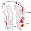
Body Part Excretory ducts in anterior part
Image Use creative commons This media file is licensed under the Creative Commons Attribution-ShareAlike License - Version 3.0.
Copyright © Klaus Rohde
Attached to Group Schizochoerus liguloideus: view page image collection
Title ligul3.gif
Image Type Drawing/Painting
Image Content Body Parts
ID 3417
3436
Scientific Name Multicotyle purvisi
Comments Growth of ventral disc of Multicotyle purvisi. Note the change in the relative length of the ventral disc during growth. The disc shows at first negative allometric growth (allometric exponent less than 1), then positive allometric growth (allometric exponent greater than 1), and finally isometric growth (allometric exponent approximately 1)
Reference redrawn from Rohde, K. (1972). The Aspidogastrea, especially Multicotyle purvisi Dawes, 1941. Advances in Parasitology 10, 77-151.
Image Use creative commons This media file is licensed under the Creative Commons Attribution-ShareAlike License - Version 3.0.
Copyright © Klaus Rohde
Attached to Group Aspidogastrea: view page image collection
Title fig23as.gif
Image Type Diagram
ID 3436
3530
Comments Note accessory seminal receptacle consisting of basal narrow and distal widened part.
Reference Redrawn from Dubinina, M.N. (1982). Parasitic worms of the class Amphilinida (Platyhelminthes). "Nauka", Leningrad.
Acknowledgements According to Dönges and Harder (1966)
Body Part Note accessory seminal receptacle consisting of basal narrow and distal widened part.
Image Use creative commons This media file is licensed under the Creative Commons Attribution-ShareAlike License - Version 3.0.
Copyright © Klaus Rohde
Attached to Group Schizochoerus africanus: view page image collection
Title afri1.gif
Image Type Drawing/Painting
Image Content Specimen(s)
ID 3530
3584
Scientific Name Cerithium moniliferum and Lobatostoma manteri
Comments Section through adhesive disc (L) of Lobatostoma manteri attached to snail tissue. Note dense fibrous tissue (F) close to adhesive disc.
Image Use creative commons This media file is licensed under the Creative Commons Attribution-ShareAlike License - Version 3.0.
Copyright © Klaus Rohde
Attached to Group Aspidogastrea: view page image collection
Cerithioidea (Caenogastropoda): view page image collection
Title fig35as.gif
Image Type Photograph
Image Content Body Parts
ID 3584
3655
Comments Note mucus exuded into water through stalk of egg, and larval hooks observed in vivo acting antagonistically in the directions indicated by the arrows
Reference Dubinina, M.N. (1982). Parasitic worms of the class Amphilinida (Platyhelminthes). "Nauka", Leningrad (in Russian).
Acknowledgements According to Dubinina (1974)
Life Cycle Stage Fully developed larva
Copyright © 1982 Nauka Publishers
Image Use ToL use only
Attached to Group Amphilina foliacea: view page image collection
Title foliac7.gif
Image Type Drawing/Painting
Image Content Specimen(s)
ID 3655
3716
Reference redrawn from Rohde, K. and Watson, N. (1988). Development of the protonephridia of Austramphilina elongata. Parasitology Research 74, 255-261.
Life Cycle Stage larva
Body Part structure of protonephridium (A and C, C is a cross-section through the proximal canal), and development of flame bulb (B is a slightly earlier stage than G. D, E and F are cross-sections through the parts indicated by arrows)
Image Use creative commons This media file is licensed under the Creative Commons Attribution-ShareAlike License - Version 3.0.
Copyright © Klaus Rohde
Attached to Group Gigantolina elongata: view page image collection
Title fig13am.gif
Image Type Diagram
Image Content Body Parts
ID 3716
3838
Scientific Name Multicotyle purvisi
Reference redrawn from Rohde, K. (1972). The Aspidogastrea, especially Multicotyle purvisi Dawes, 1941. Advances in Parasitology 10, 77-151.
Life Cycle Stage larva
Body Part nervous system
Image Use creative commons This media file is licensed under the Creative Commons Attribution-ShareAlike License - Version 3.0.
Copyright © Klaus Rohde
Attached to Group Aspidogastrea: view page image collection
Title fig08as.gif
Image Type Diagram
Image Content Specimen(s)
ID 3838
3906
Scientific Name Multicotyle purvisi
Reference redrawn from Rohde, K. (1972). The Aspidogastrea, especially Multicotyle purvisi Dawes, 1941. Advances in Parasitology 10, 77-151.
Life Cycle Stage Juveniles
Body Part with well developed ventral discs and reproductive systems
Image Use creative commons This media file is licensed under the Creative Commons Attribution-ShareAlike License - Version 3.0.
Copyright © Klaus Rohde
Attached to Group Aspidogastrea: view page image collection
Title fig21as.gif
Image Type Diagram
Image Content Specimen(s)
ID 3906
3993
Reference redrawn from Dubinina, M.N. (1982). Parasitic worms of the class Amphilinida (Platyhelminthes). "Nauka", Leningrad.
Body Part Anterior end
Image Use creative commons This media file is licensed under the Creative Commons Attribution-ShareAlike License - Version 3.0.
Copyright © Klaus Rohde
Attached to Group Gigantolina magna: view page image collection
Title magna1.gif
Image Type Drawing/Painting
Image Content Body Parts
ID 3993
Please note: Most images and other media displayed on the Tree of Life web site are protected by copyright, and the ToL cannot act as an agent for their distribution. If you would like to use any of these materials for your own projects, you need to ask the copyright owner(s) for permission. For additional information, please refer to the ToL Copyright Policies.

Contribute to the ToL

ToL Contributions

Ways to Contribute

Use of Contributions

Scientific Content

Building Treehouses

Images/Media

  go to the Tree of Life home page
top